カテゴリー別アーカイブ: English Edition

Effects of Shiatsu to the Plantar Region on Center of Gravity Sway (Part 1)

Koichi Hoshino,Munetaka Hibi
Japan Shiatsu CollegeShiatsu Department
Hiroyuki Ishizuka
Japan Shiatsu CollegeShiatsu instructor

Abstract : There are few studies on the effects of plantar region shiatsu treatment on the locomotor system. In this study, we examined the effects of shiatsu stimulation of the plantar region on standing balance, based on measurements obtained using a stabilograph. Four healthy subjects received shiatsu treatment to the plantar region for 1 min 42 sec per session. Results showed no significant differences between the stimulation group and the non-stimulation group with respect to total trajectory length, outer circumference area, rectangle area, or effective value area. Further research is required using different test subjects and research methodology.

Keywords: Shiatsu, plantar region, variance of center of gravity, balance in the upright position


I.Introduction

In shiatsu therapy, the plantar region is approached based on a variety of interpretations depending on the symptoms involved, with numerous accounts of its perceived efficacy based on experience.

However, few studies have been conducted on the effects on the locomotor system of Japanese manual therapies to this region.

In this study, as an initial step in clarifying the effects of shiatsu therapy on the muscles and structure of the plantar region and the consequent effect on locomotor function, we progressed to the pre-experimental stage in the measurement and analysis of changes to standing balance using a stabilograph. This is an interim report on the results obtained and summary of our ongoing research.

Ⅱ.Methods

1.Subjects

Research was conducted on four healthy males (mean age: 30 ± 10.68 years old) who were students at the Japan Shiatsu College. Test procedures were fully explained to each test subject and their consent obtained.

2.Test location and period

Testing was conducted in the lounge space at Japan Shiatsu College between January 29 and February 5, 2015.

3.Measurement procedure

Center of gravity sway was measured using a stabilograph (Gravicorder GS-10 Type C; Anima Corp.). Each measurement was recorded for 1 minute while subjects stood with the medial borders of their feet together, arms crossed over their chests, and eyes closed. Results were obtained for 10 measurement criteria (total trajectory length, unit trajectory length, unit area trajectory length, outer circumference area, rectangle area, effective value area, sway mean center deviation X-axis, sway center deviation X-axis, sway mean center deviation Y-axis, and sway center deviation Y-axis).

4.Stimulation

(1)Area stimulated

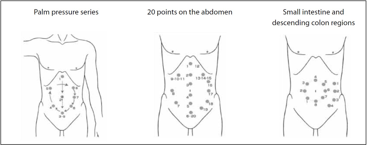
In accordance with the basic treatment points employed in Namikoshi shiatsu, 4 points were stimulated between Point 1, located in the plantar region between the bases of the second and third digits, and the edge of the heel, using 2-thumb pressure with the test subject in the prone position (Fig. 1).

4 points of plantar region
Fig. 1. 4 points of plantar region

(2)Duration and method of stimulation

Pressure was applied for 3 seconds to each of the 4 points, repeated 3 times, then single-point pressure was applied to Point 3 for 3 seconds, repeated 3 times, with a total duration of approx. 1 min 42 sec for both feet. Treatment was applied using standard pressure (pressure gradually increased, sustained, and gradually decreased), with pressure regulated so as to be pleasurable for the test subject (standard pressure) 1.

5.Test procedure

In order to average the test subjects’ learned behavior, they were randomly divided into two groups of two, Group A and Group B. Group A was scheduled to act as the non-stimulation group first, then as the stimulation group, while Group B acted first as the stimulation group, then as the non-stimulation group (Fig. 2).

Test schedule and learned behavior averaging
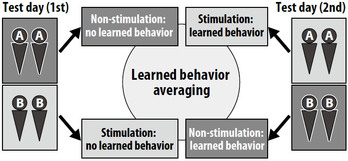
Fig. 2. Test schedule and learned behavior averaging

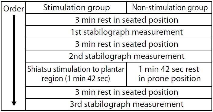
(1)Stimulation group  

The procedure was performed as follows:
1) 3 min rest in seated position
2) 1st stabilograph measurement
3) 3 min rest in seated position
4) 2nd stabilograph measurement
5) Shiatsu stimulation to plantar region
6) 3 min rest in seated position
7) 3rd stabilograph measurement

(2)Non-stimulation group

The procedure was performed as follows:
1) 3 min rest in seated position
2) 1st stabilograph measurement
3) 3 min rest in seated position
4) 2nd stabilograph measurement
5) 1 min 42 sec rest in prone position
6) 3 min rest in seated position
7) 3rd stabilograph measurement

Test procedure

6.Statistical processing

Of the data obtained from the stabilograph, measurements for total trajectory length, outer circumference area, rectangle area, and effective value area were compared between the non-stimulation group and the stimulation group by subjecting data on change rates between the 2nd and 3rd stabilograph measurements to t-testing.

Ⅲ.Results

1.Total trajectory length (Fig. 3)

Compared to the non-stimulation group, which had a change rate of 85.5 ± 7.2% (mean ± SE), the stimulation group had a change rate of 92.9 ± 7.0%, which was not statistically significant (p<0.595).

Total trajectory length
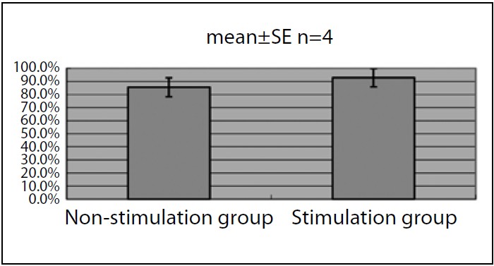
Fig. 3. Total trajectory length

2.Outer circumference area (Fig. 4)

Compared to the non-stimulation group, which had a change rate of 90.6 ± 17.8%, the stimulation group had a change rate of 79.6 ± 13.3%, which was not statistically significant (p<0.744).

Outer circumference area
Fig. 4. Outer circumference area

3.Rectangle area (Fig. 5)

Compared to the non-stimulation group, which had a change rate of 79.3 ± 14.9%, the stimulation group had a change rate of 79.4 ± 13.9%, which was not statistically significant (p<0.996).

Rectangle area
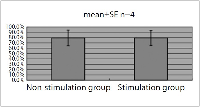
Fig. 5. Rectangle area

4.Effective value area (Fig. 6)

Compared to the non-stimulation group, which had a change rate of 92.0 ± 23.8%, the stimulation group had a change rate of 90.6 ±17.6%, which was not statistically significant (p<0.975).

Effective value area
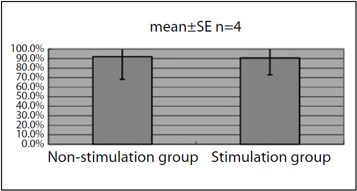
Fig. 6. Effective value area

Ⅳ.Discussion

The purpose of this study was to examine the effect of shiatsu stimulation to the plantar region on standing balance. This was based on the hypothesis that shiatsu stimulation would have a similar effect to that reported in existing research showing the effect on standing balance of sensory stimulation to the plantar region 2~6.

In this study, which was still at the preexperimental stage, we were not able to obtain data or statistical results to substantiate the effect on balance of shiatsu to the plantar region. However, each sample observed suggested a trend in the effect of shiatsu stimulation, so there is a chance that a different result will be obtained when testing is conducted with a larger sample size.

On the other hand, all measurement values obtained from this sample were from healthy test subjects considered to be within the standard range 7. It was assumed that, for test subjects within such a range, measurement values would be easily variable based on physical condition, a factor which cannot be averaged out through uniform test procedure. For this reason, it is difficult to fully investigate the effect of shiatsu stimulation on standing balance using the measurement data obtained from a stabilograph alone.

In future study, it will be necessary to consider an experimental method that includes examination of test subjects and the use of other measurement criteria in addition to the stabilograph, with integrated analysis of the results obtained.

Ⅴ.Conclusion

Shiatsu stimulation of the plantar region in four healthy test subjects did not produce a statistically significant change in measurement values using a stabilograph.

Testing of the effect of plantar region shiatsu on standing balance was insufficient due to the limitations of the test procedure employed at this stage, necessitating a reexamination of the methods employed in testing.

VI.References

1. Ishizuka H et al: Shiatsu ryohogaku: 96, International Medical Publishers, Ltd. Tokyo, 2008 (in Japanese)
2. Okubo J et al: Sokuseki atsujuyouki ga jushin doyo ni oyobosu eikyo ni tsuite. Jibirinsho 72: 1553-1562, 1979 (in Japanese)
3. Ito A et al: Sokutei sokuakkaku ga ritsui shisei no jushin doyo ni ataeru eikyo. Nihon rigaku ryoho gakujutsu taikai 2004: A1113-A1113, 2005 (in Japanese)
4. Utsunomiya Y et al: Kankaku shigeki ga seiteki ritsui ni oyobosu eikyo. Nihon rigaku ryoho gakujutsu taikai 2005: A0853-A0853, 2006 (in Japanese)
5. Kamei S et al: Sokutei no kankaku shigeki ga jushin doyo ni ataeru eikyo ni tsuite. Aino gakuin kiyo 20: 27-40, 2006 (in Japanese)
6. Nose T et al: Boshi sokuteibu he no shokuatsu shigeki ga shisei seigyo ni oyobosu eikyo. Nihon rigaku ryoho gakujutsu taikai 2009: A4P2047-A4P2047, 2010 (in Japanese)
7. Imamura K et al: Jushin doyo kensa ni okeru kenjosha data no shukei. Equilibrium research, supplement 12: 15-23, 1997 (in Japanese)


Measurement of the Psychological Effect of Full Body Shiatsu Therapy: a Case Report

Shinpei Oki
Representative, Nekonote Shiatsu

Abstract : This report examines the case of a female patient in her 20s who received three full body shiatsu treatments between May 24 and June 7 2015, with the objective of reducing psychological stress. The psychological effect was evaluated using the Profile of Mood States (POMS) index. Following treatment, improvements in the t-scores of all six POMS factors were observed. This suggests that full body shiatsu therapy has a stress-relief effect, which may be verified through further studies.

Keywords: shiatsu, stress, POMS


I.Introduction

 So many people are feeling the effects of psychological stress in modern society that stress can be considered endemic 1. In Japan, many people look to alternative therapies, including anma, massage, and shiatsu, for treatment of stress.

 Multiple studies have been conducted into the effects of manual therapy on stress relief 2~5, confirming its effectiveness. Research has also been conducted into the use of shiatsu for treating stress 6, but insufficient data exists on the effects of general shiatsu carried out by a therapist on a patient. In this paper, we report on a case in which full body shiatsu used to alleviate stress with psychological stress measured using a mood profile, which will serve as a springboard for future investigation.

Ⅱ.Methods

Test subject

 Female office worker in her 20s

Period

 May 24 to June 7, 2015 (3 sessions)

Location

 Patient’s home

Treatment method

 Namikoshi-style full body shiatsu, starting in lateral position

Evaluation method

 In order to evaluate psychological effects, a Japanese-language POMS™ test (Kaneko Shobo) was administered immediately before and after treatment. POMS is a mood profile test developed by McNair et al in the U.S., which employs answers to 65 questions to enable simultaneous measurement of six factors: tension-anxiety, depression, anger-hostility, vigor, fatigue, and confusion 7. The POMS results for this report were converted from raw data to t-scores and totaled. POMS has been implemented on large groups of healthy adult males and females and standardized, with t-scores calculated for mean value and standard deviation by age and sex. The t-score is calculated as 50 + 10 x (raw score – average score) / standard deviation. If the raw score is equal to the average score, the t-score will be 50. The lower the t-score, the lower the tension-anxiety, depression, anger-hostility, vigor, fatigue, or confusion. Thus, for vigor, a higher t-score indicates a more favorable condition 7.

 The goal and measurement procedure for POMS was fully explained to the patient and her consent obtained.

Ⅲ.Results

History of present illness

 The patient was transferred to a new department at work in April 2015 and, still unaccustomed to the new workplace and job responsibilities, was experiencing high daily stress levels. Work mainly involved VDT (video display terminal) operation, with over 7 hours per day spent engaging in computer input.

Medical history

 Inguinal hernia (surgery completed in 2013)

Family history

 No relevant items

Subjective findings

  • Sleep disorder
    On some days, the patient had difficulty getting to sleep because she was unable to relax emotionally. The harder she tried to sleep, the more difficult it would become.
  • Neck, shoulder, and lumbar pain
    The patient experienced chronic neck and shoulder stiffness. Perhaps because she assumed the same posture for extended periods, she experienced a grinding pain when she extended her back.

Examination findings

  • Observation
    The patient’s complexion was poor, with bags under her eyes and numerous pimples around her jaw. Head-forward poster with exaggerated lumbar kyphosis was observed.
  • Palpation
    Cervical region: Hypertonus was confirmed in anterior and middle scalenus, splenius capitis, rectus capitis posterior major and minor, and semispinalis capitis. Misalignment of the lower cervical vertebrae was also observed.
    Shoulder, dorsal, and lumbar regions: Hypertonus was confirmed in the upper trapezius, levator scapulae, and quadratus lumborum.
    Abdomen: The lower abdomen was flaccid and induration was observed in the descending colon region (left umbilical region).

Treatment #1 (May 24, 2015)

  • Rigidity in the dorsal region was alleviated.
  • Post-treatment, the patient reported fullbody relaxation and mild drowsiness.

Treatment #2 (May 30, 2015)

  • Patient reported that she slept well after the previous treatment and that she awoke the next morning with no feelings of lethargy.
  • She also stated that her neck and shoulders felt lighter than usual.

Treatment #3 (June 7, 2015)

  • Patient reported that she slept well for several days after treatment and that she felt comparatively fresh on waking.
  • She still felt stiffness in the neck and shoulders, but it was not severe. Her lumbar region was still slightly stiff, but not painfully so.
  • She felt that her stress level was lower as well.

 Table 1 shows the POMS t-scores measured before and after all three treatments. Aside from the anxiety factor on May 24 and the vigor factor on May 30, the values for all factors showed improvement posttreatment, with a general trend toward improvement as the treatments progressed (Fig. 1).

Table 1. T-scores for six POMS factors

Fig. 1. Changes in t-scores for six POMS factors

Ⅳ.Discussion

 In the case presented in this report, the patient showed improvement in all six POMS factors over the course of three treatments. Kamohara et al and Asai et al demonstrated the possibility for suppression of sympathetic nervous system activity using shiatsu to the abdominal region and the dorsal region, respectively 8-9. Also, Yokota, Watanabe, and Tadaka et al reported miotic (pupil contraction) response to shiatsu to the anterior cervical, lower leg, sacral, and head regions, respectively, possibly due to either suppression of the sympathetic nervous system or stimulation of the parasympathetic nervous system 10~12. The patient in this case report received full body shiatsu, including comprehensive shiatsu stimulation to all of the above-mentioned regions, so it is probable that a relaxation effect was achieved due to both suppression of the sympathetic nervous system and stimulation of the parasympathetic nervous system. In addition, Kato reported that, in restraint-stressed rats, acupuncture electrostimulation lead to normalization of secretion of monamines including dopamine and serotonin in all areas of the brain 13, so one might consider the possibility that a similar mechanism occurs with shiatsu stimulation as well.

 A single case such as this is insufficient evidence to argue for the effectiveness of shiatsu therapy for treatment of stress. Verification of the effectiveness of shiatsu as a means of stress alleviation will require a study employing statistical methodology, which I hope to pursue as a research topic in the future.

Ⅴ.Conclusion

 Improvement was observed in all six POMS factors over the course of three full body shiatsu treatments.

References

1. Govt. of Japan Cabinet Office website: Heisei 20 nendo-ban kokumin seikatsu akusho, 2008 (in Japanese)
2. Kober A, Scheck T, et al: Auricular acupressure as a treatment for anxiety i prehospital transport setting. Anesthesiology 98: 1328-1332, 2003
3. Sato T: Kenko na seijin josei ni okeru hando massaji no jiritsu shinkei katsudo oyobi kibun he no eikyo. Yamanashi daigaku kango gakkaishi 4(2): 25-32, 2006(in Japanese)
4. Fujita K: Haibu massaji ni yoru seijin dansei no shintaiteki • sinnriteki eikyo. Ube furontia diagaku kangogaku janaru 4(1): 37-43, 2011 (in Japanese)
5. Sakai K et al: Kenko na josei ni taisuru takutiru kea no seiriteki • shinriteki koka. Nippon kango kenkyu gakkaishi 35(1): 145-152, 2012 (in Japanese)
6. Honda Y et al: Serufu keiraku shiatsu ga kibun ni oyobosu kyusei koka to sono yuzabiriti ni kan suru kenkyu. Kenko Shien 15(1): 49-54, 2013 (in Japanese)
7. Yokoyama K: Nihongoban POMS tebiki, 1-7. Kaneko Shobo, Tokyo, 1994 (in Japanese)
8. Kamohara H et al: Effects of Shiatsu Stimulation on Peripheral Circulation. Toyo ryoho gakko kyokaishi(24): 51-56, 2002 (in Japanese)
9. Asai S et al: Effects of Shiatsu StimuIation on Muscle PIiability. Toyo ryoho gakko kyokaishi (25): 125-129, 2001 (in Japanese)
10. Yokota M et al: Effect on Pupil Diameter of Shiatsu Stimulation to the Anterior Cervical and Lateral Crural Regions. Toyo ryoho gakko kyokaishi (35): 77-80, 2011 (in Japanese)
11. Watanabe T et al: Effect on Pupil Diameter,Pulse Rate, and Blood Pressure of Shiatsu Stimulation to the Sacral Region. Toyo ryoho gakko kyokaishi (36): 15-19, 2012 (in Japanese)
12. Tadaka S et al: Tobu he no shiatsu shigeki ga doko chokkei • myakuhakusu• ketsuatsu ni oyobosu koka. Toyo ryoho gakko kyokaishi (37): 154-158, 2013 (in Japanese)
13. Kato M: Kosoku sutoresu ratto he no hari tsuden shigeki no nonai monoamin ni oyobosu eikyo. Meiji shinkyu igaku (27): 27-45, 2000 (in Japanese)


Shiatsu Therapy for a Patient with Suspected Peripheral Neuropathy while Diagnosed with Traumatic Cervical Spinal Cord Injury

Ichiro Maruyama
Graduated Japan Shiatsu College in 2012

Abstract : This report examines the case of a patient diagnosed with traumatic cervical spinal cord injury and suspected peripheral neuropathy (flaccid paralysis of the lower extremities) who was treated with shiatsu therapy for the alleviation of dorsal muscle tension. After 29 treatments, lower-limb motor function recovered. This suggests that hypertonicity in paraspinal muscles was significantly related to the motor dysfunction due to peripheral neuropathy. Considering other reports on the effect of shiatsu stimulation in improvement of muscle pliability, we conclude that in this patient the decrease in muscle hypertonicity due to shiatsu therapy resulted in improved blood circulation and increased spinal range of motion, leading to a recovery of motor function.

Keywords: flaccid paralysis of the lower extremities, shiatsu therapy, dorsal muscle tension


I.Introduction

 Spinal cord injury refers to injury of the spinal cord where it is protected within the spinal canal. Depending on the level of the spinal cord injury, symptoms presented may include motor, respiratory, circulatory, urinary, digestive, or other dysfunctions. Treatment is divided between initial phase treatment and chronic phase treatment, with initial phase treatment including pharmacotherapy, localized rest, cranial traction, and surgery, while chronic phase treatment centers on rehabilitation. Here, we report on a case in which the symptoms of a patient diagnosed with traumatic cervical spinal cord injury virtually disappeared following therapy.

Ⅱ.Methods

Location

 Patient’s home

Period

 August 25 to December 1, 2014 (Number of treatments: 29)

Test subject

 82 year old female

History of present illness

 The patient sustained a traumatic cervical spinal cord injury 46 years previously. Rehabilitation restored motor function in the upper limbs, but paralysis (paraplegia) of the lower limbs remained and she had been confined to a wheelchair ever since. Six years previously she sustained a fracture to her right humerus, and later required amputation of the arm due to pyogenic osteomyelitis. Two years previously she was diagnosed with tuberculosis and admitted to a tuberculosis ward, after which she became bedridden. After discharge from the hospital, she developed pain in her upper limb and dorsal regions, and it was arranged for her to received homecare massage for alleviation of the pain.

Medical history

 Paraplegia (circulatory organ, urinary, and digestive organ dysfunction) due to spinal cord injury; gallbladder cancer; pancreatic cancer; tuberculosis; amputation of right arm due to pyogenic osteomyelitis

Treatment

  • Shiatsu to cervical, dorsal, sacral, and gluteal regions in lateral position
  • Shiatsu to left upper limb and lower limbs in supine position (emphasis on treatment of lower limbs)

Evaluation

  • Pain evaluated using 10-step VAS
  • Manual muscle testing (MMT)

III.Results

August 25 (Treatment #1)
Pre-treatment findings
 Subjective findings

  • Motor paralysis and sensory dysfunction inferior to lumbar region
  • Numbness below knees
  • Bladder and rectal dysfunction
  • Pain in upper limb and dorsal regions
  • Hot and cold flashes (excessive sweating from neck up)

 Objective findings

  • Limited range of motion in left shoulder joint
  • Flaccid paralysis and sensory dysfunction of lower limbs
  • Pain in dorsal and gluteal regions

 Post-treatment findings

  • Hot and cold flashes alleviated due to improved circulation
  • Pain reduced

September 4 (Treatment #4)
 Post-treatment findings

  • Dorsal region muscle tension reduced (thoracolumbar junction)
  • Pain in medial femoral region absent
  • Slight return of sensory function in femoral region (femoral nerve, obturator nerve)
  • Muscle contraction observed in femoralregion (adductor muscles)

September 8 (Treatment #5)
 Post-treatment findings

  • Plantar pain absent
  • Patient found shiatsu to sacral region pleasurable

September 18 (Treatment #8)
 Post-treatment findings

  • Patient felt urinary and bowel sensations (improvement of bladder and rectal dysfunction)
  • Return of sensory function to femoral region

October 2 (Treatment #12)
 Post-treatment findings

  • Muscle contraction observed in femoral region (femoral nerve, obturator nerve)

October 30 (Treatment #20)
 Post-treatment findings

  • Muscle contraction observed in femoral region (sciatic nerve)

November 6 (Treatment #22)
 Post-treatment findings

  • Movement observed in hip joint (flexion, extension, external rotation, internal rotation)
  • Movement observed in knee joint (flexion, extension)
  • Left shoulder joint more stable; pain absent
  • Changed sensation distal to knee

November 17 (Treatment #25)
 Post-treatment findings

  • Movement observed in ankle joint and toes (flexion, extension) with patient lying in lateral position
  • Patient able to form slight bridge (elevation of gluteal region)

December 1 (Treatment #29)
 Post-treatment findings

 Subjective findings

  • Patient experiences numbness in calcaneal region
  • Pain eliminated

 Objective findings

  • Return of motor function inferior to lumbar region
  • Improvement to bladder and rectal dysfunction

Table 1. 10-step VAS pain scale values (post-treatment)

Table 2. Manual muscle testing (MMT) of lower limbs

IV.Discussion

 In most cases of spinal cord injury, the vertebrae undergo dislocation fracture due to an external force, with concomitant damage to the spinal cord. Characteristics vary depending on the level and degree of spinal cord injury (complete or incomplete paralysis), but immediately after the injury spinal shock occurs and autonomy is lost in the spinal cord inferior to the injury. Specifically, flaccid paralysis occurs, with loss of all motor, sensory, and deep tendon reflex function, while at the same time autonomic nervous function is also impaired. Following the recovery period, reflex functions in the spinal cord inferior to the injury are recovered, resulting in spastic paralysis, characterized by hyperreflexia of the deep tendon reflexes 1.
 In this case, since the patient exhibited flaccid paralysis from post-injury to the present, it is likely that this was a case not of spinal cord injury, but rather of spinal cord compression. In other words, assuming lower motor neuron damage and comparing spinal cord injury level with ADL levels, since T1 ADLs (upper limbs normal, full wheelchair mobility) were possible and T6 functions (circulatory organ stability) were unstable, it was determined that there was an irregularity in the upper thoracic vertebrae. Clinical findings indicated that the thoracic spine was straight, with almost no curve in the thoracic vertebrae. We may hypothesize that this caused hypertonus in the dorsal musculature, causing lower motor neuron damage, pain, and motor dysfunction.
 Based on the above determination of peripheral neuropathy due to spinal cord compression, shiatsu therapy was carried out with the objective of alleviating pain and restoring motor function in the patient. As a result, after 29 treatments, decrease in VAS values as an indicator of pain (Table 1) and recovery of muscle strength as determined by manual muscle testing (Table 2) were observed, although numbness remained in the calcaneal region. If this were a case of spinal cord injury, such rapid return of function would be unlikely 2-3. It is therefore reasonable to assume that recovery was due to shiatsu treatment of peripheral neuropathy caused by nerve entrapment due to hypertonic muscles.
 At the very least, in this case it is highly likely that hypertonicity in paraspinal muscles was significantly related to the motor dysfunction due to peripheral neuropathy. Considering other reports on the effect of shiatsu stimulation in improvement of muscle pliability 4~6, we conclude that in this patient the decrease in muscle hypertonicity due to shiatsu therapy resulted in improved blood circulation and increased spinal range of motion, leading to a recovery of motor function.

V.Conclusion

 Even in patients afflicted by long-term peripheral neuropathy (pain and motor dysfunction), recovery through shiatsu therapy is possible.

VI.References

1. Nara N et al: Toyo ryoho gakko kyokai rinsho igakukakuron (dai 2 han) sekizui sonsho. Ishiyaku shuppan KK: 171-173, 2010 (in Japanese)
2. Shinno Y: Massho shinkei shogai no rihabiriteshon. Nihon rihabiriteshon igakukaishi 28 (6): 453-458 (in Japanese)
3. Nishiwaki K et al: Massho shinkei sonshogo no shinkeisaisei to rihabiriteshon. Nihon rihabiriteshon igakukaishi 39 (5): 257-266, 2002 (in Japanese)
4. Asai S et al: Effects of Shiatsu Stimulation on Muscle Pliability. Toyo ryoho gakko kyokai gakkaishi (25): 125-129, 2001 (in Japanese)
5. Sugata N et al: Effects of Shiatsu Stimulation on Muscle Pliability(Part2). Toyo ryoho gakko kyokai gakkaishi (26): 35-39, 2002 (in Japanese)
6. Eto T et al: Effects of Shiatsu Stimulation on Muscle Pliability(Part3). Toyo ryoho gakko kyokai gakkaishi (27): 97-100, 2003 (in Japanese)


A Case of Posture Correction with a Combination of Pressure Application and Mobilization

Genta Niikura
Clinic Director, Genta Chiryoin

Abstract : In clinical practice, one encounters many patients presenting subjective symptoms of shoulder stiffness or back pain. Here we examine a case in which symptoms were alleviated through posture and joint correction, in addition to using shiatsu therapy to reduce muscle tension. By combining the pressure applications of shiatsu therapy with mobilization, it was possible to achieve an effect on both muscles and joints.

Keywords: shiatsu therapy, pressure application, exercise therapy, posture correction


I.Introduction

 In clinical practice, one often encounters patients for whom, even though muscle tension is alleviated through shiatsu therapy consisting of pressure application to muscles and soft tissues, similar symptoms return after several days or weeks.

 It was our opinion that these symptoms could be more effectively treated with a combination of shiatsu therapy and ongoing posture correction and joint adjustment.

 Here, we report on a case in which significant therapeutic effect was achieved through joint adjustment and posture correction via the use of pressure applications combined with mobilization.

Ⅱ.Methods

Subject

 Female child care worker in her 30s

Location

 This clinic (Genta Chiroin)

Period

 March 30 to April 12, 2014

Primary complaint

 Work involves frequent crouching, leading to lumbar pain, stooped posture, and severe shoulder stiffness; patient told by coworkers that she has poor posture

Treatment method

 Full body shiatsu 1 combined with mobilization for shoulder, hip, and sacroiliac joints

  • For rounded back:
    Prone position: Palmar pressure to spine, spinous process adjustment
  • For internal rotation of shoulder joints:
    Lateral position:
    Pressure applications to superior angle of scapula, sub-clavicular region, and coracoid process, plus adjustment procedure to scapula
  • For Lumbar kyphosis:
    Supine position: Palmar pressure to abdomen and inguinal region
    Prone position: Adjustment of hip and sacroiliac joints
  • For posterior pelvic tilt:
    Supine position: Palmar pressure to abdomen and inguinal region
    Prone position: Adjustment of hip and sacroiliac joints

III.Results

Treatment #1 (March 30, 2014)
Pre-treatment findings
 Subjective findings

  • Work involves frequent crouching, leading to lumbar pain, stooped posture, and severe shoulder stiffness; patient told by coworkers that she has poor posture Objective findings
  • Exaggerated posterior pelvic tilt, rounded back, exaggerated internal rotation of shoulders (Fig. 1)

Post-treatment findings
 Subjective findings

  • Reduced sensations of shoulder stiffness and lumbar pain; reduced discomfort at work, even after maintaining same posture for a prolonged period

 Objective findings

  • Tension in lumbar musculature reduced due to creation of anterior pelvic tilt and lumbar lordotic curve; reduced internal rotation of shoulders due to improved shoulder posture (Fig. 2)

treatment #1

Treatment #2 (April 12, 2014)
Pre-treatment findings
 Subjective findings

  • Patient told by those around her that her posture had improved; alleviation of lumber pain

 Objective findings

  • Anterior pelvic tilt maintained; exaggerated internal rotation of shoulders observed (Fig. 3)

Post-treatment findings
 Subjective findings

  • Alleviation of symptoms of shoulder stiffness and lumbar pain; reduced discomfort, even after maintaining same posture for a prolonged period

 Objective findings

  • Improvement of exaggerated internal rotation of shoulders (Fig. 4); alleviation of muscle tension due to adjustment of joint position

treatment #2

IV.Discussion

 According to the Comprehensive Survey of Living Conditions by the Japanese Ministry of Health, Labour and Welfare 2, the two most common symptoms experienced by both men and women in Japan are, in order, stiff shoulders and lumbar pain. Little has changed in this situation, and a comparatively large number of patients visiting our clinic list stiff shoulders or lumbar pain as their primary complaint.

 It is my experience in a clinical setting that, in order to alleviate shoulder stiffness or lumbar pain, effects are longer lasting if reduction of excess muscle tension is used in combination with joint adjustment.

 The reason is that, as humans are bipedal, they must continually maintain posture in opposition to gravity. The extensors, trunk muscles, and other antigravity muscles must maintain contraction in order to resist gravity and maintain proper posture, which when disrupted is corrected by postural reflexes 3. It follows that a greater load is placed on these muscles when proper posture and skeletal alignment are regularly disrupted during routine daily activities. For this reason, correction of chronically disrupted postural and skeletal alignment should help alleviate symptoms of shoulder stiffness and lumbar pain.

 In this case, initial examination revealed marked postural disruption from the line of gravity (Fig. 1), indicating probable hypertonus in the pectoralis major and other shoulder internal rotator muscles along with reduced tonus of the antigravity muscles. Also, hypertension in the gluteus maximus and hamstring muscles were likely responsible for the posterior pelvic tilt.

 In the initial treatment, hypertonus in the gluteus maximus and hamstrings was improved, along with overextension of the quadratus lumborum and erector spinae muscles, as was evidenced by the reductions in posterior pelvic tilt and lumbar kyphosis. Also, concerning the shoulder joints, changes to the position of the scapula were likely due to reduced hypertonus in the internal rotators, including the pectoralis major, latissimus dorsi, and subscapularis (Fig. 2).

 Prior to treatment #2, a slight internal rotation of the shoulder joints was observed, though no major postural disruption from the line of gravity compared to post-treatment #1 was apparent (Figs. 2, 3). In treatment #2, further improvements to pliability in hypertoned muscles improved balance with over-extended muscles, causing positional changes to the shoulder joint and humerus that likely resulted in reduced tension in the trapezius, sternocleidomastoid, and other neck muscles that led to improvements in head position.

 It is difficult to determine whether disrupted posture due to routine daily activities led to muscular hypertonus and hypotonus or whether the problem was due to irregularities in joint alignment, but since multiple reports have shown that shiatsu stimulation effectively increases muscle pliability 4-6, in this case it is likely that pressure application relaxed hypertoned muscles that were the cause of postural disruption while mobilization improved joint positioning, resulting in alleviation of symptoms.

V.Conclusions

 When the pressure applications of shiatsu therapy are combined with mobilization, there is a tendency for symptoms of stiff shoulders and lumbar pain to be alleviated due to elimination of muscle hypertension and improved joint positioning. However, because this report only contains one example, it will be necessary to study a larger number of cases.

VI.References

1. Ishizuka H: Shiatsu ryohogaku, first revised edition, International Medical Publishers, Ltd. Tokyo, 2008 (in Japanese)
2) 2. Japanese Ministry of Health, Labour and Welfare: Kokumin seikatsu kiso chosa. 2013, http://www.mhlw.go.jp/toukei/list/20-21.html (in Japanese)
3. Toyo ryoho gakko kyokai: Seirigaku. Ishiyaku shuppan KK, 1990 (in Japanese)
4. Asai S et al: Effects of Shiatsu Stimulation on Muscle Pliability. Toyo ryoho gakko kyokai gakkaishi (25): 125-129, 2001 (in Japanese)
5. Sugata N et al: Effects of Shiatsu Stimulation on Muscle Pliability(Part2). Toyo ryoho gakko kyokai gakkaishi (26): 35-39, 2002 (in Japanese)
6. Eto T et al: Effects of Shiatsu Stimulation on Muscle Pliability(Part3). Toyo ryoho gakko kyokai gakkaishi (27): 97-100, 2003 (in Japanese)


Effects of Inguinal Region Shiatsu on Walking Ability

Hiroki Koizumi
Shiatsu Department, Japan Shiatsu College
Yasutaka Kaneko
Shiatsu instructor, Japan Shiatsu College; Clinic director, MTA Shiatsu Chiryoin

Abstract : The Timed Up and Go (TUG) test was employed to determine the effect of inguinal region shiatsu on walking ability. The post-treatment time was shorter than the pre-treatment time, suggesting that shiatsu stimulation may improve walking ability at least temporarily.

Keywords: Timed Up and Go Test, iliopsoas muscle, inguinal region, shiatsu


I.Introduction

 Yoshinari et al have reported on the possibility that shiatsu stimulation to the inguinal region increases range of motion for hip extension and lumbar vertebrae retroflexion in standing automatic trunk retroflexion 1, postulating that shiatsu stimulation to the inguinal region reduced tension in the iliopsoas muscles, increasing range of motion in the lumbar vertebrae and hip joints. However, there was no reference to functional changes. In this comparatively simple study, we observe changes to walking ability after inguinal region shiatsu using the highly reliable TUG test as an evaluative tool. The TUG test, devised by Podsiadlo et al in 1991 2, is a widely used evaluation index for walking ability in the elderly.

Ⅱ.Methods

Location

 8th floor classroom, Namikoshi Institute • Japan Shiatsu College

Test subject

 66-year-old male (no history of central nervous system dysfunction, bone fractures, muscle rupture, degenerative arthritis, or other disorders that may affect lower limb function)

Period

 September 4, 12, 19, October 3, 2015 (4 treatments over 30 days)

Stimulation

 The subject was placed in a relaxed supine position with all four limbs extended. The therapist stimulated three points over the inguinal ligament, extending medioinferiorly from the anterior superior iliac spine to the lateral border of the pubic bone 3. Stimulation consisted of (1) palmar pressure (pressure using the thenar eminence) and (2) shiatsu using the therapist’s thumbs of both hands, held for approximately 5 seconds per point, applied for 5 minutes each on the left and right sides. Strength of pressure was such that, when the therapist’s palm and thumbs sank into the skin and subcutaneous tissue, he was able to feel the inguinal ligament and femoral pulse, at a pressure that was comfortable for the subject.

Evaluation

 The TUG test was used to evaluate pre- and post-treatment times required. An armless chair was used, with a red cone placed 3 meters directly in front of the leading edge of the front leg as a marker. Responding to a verbal signal, the subject was required to stand up from the chair, walk around the cone, and return to sit in the chair. The time required to complete this task was measured with a stopwatch. The task was performed immediately before and immediately after stimulation, (1) at regular walking speed; and (2) at maximum walking speed, once for each. The times were measured and the time for walking speed at (2) maximum effort was used as the measurement value.

III.Results

 Post-stimulation times were reduced compared to pre-stimulation for all four sessions. No overall time reduction was seen for pre-stimulation or post-stimulation times over the entire period. (Table 1, Fig. 1)

Table 1. TUG times (sec)

Fig. 1. TUG time changes

IV.Discussion

 The iliopsoas is comprised of two muscles, the iliacus and the psoas major, which come together in the pelvic cavity to form the iliopsoas before passing through the muscular lacuna below the inguinal ligament and inserting onto the lesser trochanter of the femur. Shiatsu of the inguinal region targets pressure to the inguinal ligament, with pressure directed more or less perpendicularly to the skin’s surface on points along the inguinal ligament from the anterior superior iliac spine to the lateral border of the pubic bone. The pressure therefore should penetrate to the iliopsoas muscle.

 When walking at a moderate pace, after reaching extension at the end of the stance phase the lower limb swings forward like a pendulum, allowing the foot to move forward without employing the iliopsoas muscle. However, in effortful walking, the iliopsoas contracts powerfully during the initial-to-mid swing phase, flexing the extended leg to swing it forward 4. According to research conducted by Anderson et al using an electromyograph, the effect of these muscles on walking is greater the faster the pace 5. It is also likely that iliopsoas functionality also plays a role in pelvic stability while walking, as well as emergency postural control when balance is lost.

 Eto reported on the probability that shiatsu stimulation improves regulation of muscle output 6, hypothesizing that this may be due to its effect on kinetic and sustained neuromuscular units along with increase in local blood supply. Similarly, the reduced post-treatment times recorded in this study were likely due to changes in the condition of the iliopsoas muscles due to shiatsu stimulation of the inguinal region, affecting their function during effortful walking to result in increased walking speed.

 Regarding the fact that no cumulative time reduction was observed during the overall test period, this was likely because shiatsu stimulation to the iliopsoas in isolation did not result in a fixed change to the condition of the muscle. We hypothesize that, in order to achieve a more lasting change, it would be necessary to effect changes in hip flexors other than the iliopsoas, including the rectus femoris and tensor fasciae latae, along with antagonists such as the gluteus maximus and hamstrings, leading to changes of alignment in the sagittal plane for the hip joint and pelvis.

 In this case, because no control was used, we cannot rule out the possibility that the reduced times were due to a learning effect in the test subject. Further research employing test methodology that includes a control is required in order to verify the effect of shiatsu stimulation in isolation.

V.Conclusion

 Shiatsu stimulation to the inguinal region resulted in a tendency for TUG times to be shorter post-stimulation than pre-stimulation.

VI.References

1. Yoshinari K et al: Effect on Spinal Mobility of Shiatsu Stimulation to the Inguinal Region. Toyo ryoho gakko kyokai gakkaishi 32: 18-22, 2008 (in Japanese)
2. Podsiadlo D, Richardson S: The timed “Up & Go”: a test of basic functional mobility for frail elderly persons. Journal of the American Geriatrics Society, 39.2: 142-148, 1991
3. Ishizuka H et al: Shiatsu ryohogaku. International Medical Publishers, Ltd. Tokyo: 102, 2008 (in Japanese)
4. Neumann D, Shimada T, Hirata S:Kinkokkakukei no kineshioroji. Ishiyaku Shuppan, Tokyo: 573-574, 2005 (in Japanese)
5. Andersson EA et al: Intramuscular EMG from the hop flexor muscles during human locomotion. Acta Physiologica Scandinavica Vol. 161, Issue 3: 361-370, 1997
6. Eto T: Shiatsu ni yoru teihaikutsuryoku no henka ni tsuite. Nihon shiatsu gakkai (2): 10-12, 2013 (in Japanese)


The Effect of Standard Namikoshi Abdominal Region Shiatsu on Shortdistance Sprint Performance

Keisuke Okubo, Maho Nakano
Japan Shiatsu CollegeShiatsu Department
Hiroyuki Ishizuka
Japan Shiatsu CollegeShiatsu instructor

Abstract : Sports are receiving increasing attention in Japan ahead of the 2020 Tokyo Olympic Games. The goal of this study was to verify the effect of standard Namikoshi shiatsu therapy on sports performance.
After adequate warm-up, the test subject performed five 50-meter sprints separated by 5-minute rest intervals. The sprints were timed and photographed using a fixed camera to facilitate running posture analysis. Abdominal shiatsu, consisting of the standard 20 points on the abdomen, repeated 3 times, was applied before the initial run and during each 5-minute interval. During control testing, the subject spent the same time resting in supine position. Testing was performed on different days for shiatsu and control sessions.
On average, sprint times were shorter when shiatsu had been applied. Comparison of photographic images also showed changes in trunk rotation, knee flexion, and stride length. These results suggest that Namikoshi standard abdominal shiatsu consisting of pressure to 20 points on the abdomen may have positive effects on sprint performance.

Keywords: Shiatsu, run, sprint, abdominal region, abdominal pressure, trunk, exercise, time, 50m, track and field, manipulative therapy, angular motion, image, rectus abdominis, obliquus externus abdominis muscle, obliquus internus abdominis muscle, Olympic, length of stride, twist, motion, ROM, range of joint motion, joint, load, track, race, performance, massage, start, dash, run, crouch start, running motion, abdominal region shiatsu based on Namikoshi shiatsu therapy’s standard procedures


I.Introduction

 In this study, we examined the effect of standard Namikoshi 20-point abdominal shiatsu 1 applied to a runner prior to running a 50-meter sprint on ankle, knee, trunk, and shoulder joint ROM, hip flexion speed, and starting hip position, and verified the accompanying changes to stride length and run time.

 Measurements were taken after applying shiatsu stimulation to the test subject on August 15, with control measurements taken on August 2 when the test subject had received no shiatsu stimulation. Shiatsu stimulation consisted of standard Namikoshi 20-point abdominal shiatsu, repeated 3 times, before the initial run and during the 5-minute rest interval prior to each of 5 runs. Adequate stretching was carried out prior to running 2.

Ⅱ.Methods

Dates

 August 2 and 15, 2015

Location

 Komazawa Olympic Park athletic field, straight track

Test subject

 27-year-old male Weight: 52 kg Height: 161 cm
 Played soccer from elementary school to university

Equipment

 CASIO Digital Sports Stop Watch HS-70W

Imaging equipment

 SONY HANDYCAM HDR-CX420
 iPhone 6plus

Editing application

 Adobe premiere pro CC 2015
 Adobe photoshop CC 2015

Fig. 1. Experiment protocol

III.Results

vol4_06fig2

vol4_06fig3

vol4_06fig4

vol4_06fig5

vol4_06fig6

vol4_06fig7

vol4_06fig8

Table 1. Comparison of times (sec) with and without abdominal shiatsu

Table 2
Table 2.


IV.Discussion

 The short-distance running cycle can be divided into two phases: (1) the support phase (when the sole of the foot is in contact with the ground); and (2) the recovery phase (when the sole of the foot is not in contact with the ground). Phases (1) and (2) can each be further divided into three sub-phases. The (1) support phase includes (1)-1 foot strike (the period when a portion of the sole of the foot is in contact with the ground); (1)-2 mid support (The period from when the sole of the foot is in full contact with the ground, supporting the body’s weight, to immediately before the heel loses contact with the ground); and (1)-3 takeoff (the period from when the heel loses contact with the ground to when the toes leave the ground). The (2) recovery phase includes (2)-1 follow-through (the period from when the sole of the foot leaves the ground to when rearward motion of the lower leg ends); (2)-2 forward swing (the period when the lower leg is moving from back to front); and (2)-3 foot descent (the period immediately prior to when the sole of the foot makes contact with the ground). Specific muscles are active during each of these phases, which may vary depending on running speed. When observing the muscles active while running, the abdominal muscles are strongly active only when running 100 m at an average speed of 36 km/h, as compared to 1 km at an average speed of 12 km/h or 16 km/h. During the running cycle, strong abdominal muscle activity is observed from (1)-2 to (2)-2 3.

 The reason abdominal muscle activity is only observed in short-distance running is due to the strong angular momentum generated between arm swinging and the pelvis. While running, the pelvis produces rotational motion on a vertical axis, generating angular momentum around the vertical axis. Arm swinging is important for reducing trunk deflection due to this motion; in practice, the angular momentum produced by arm swinging eliminates the trunk deflection due to pelvic angular momentum. This is a distinguishing characteristic of short-distance running 4.

 In running there is an ideal leg trajectory. According to research conducted using a sprint training machine, leg motion effectively utilizes flexible twisting and rotational motions in the pelvis and trunk and is also necessary to adroitly maintain balance. Also important is that this motion originates in the epigastric fossa, around the level of the upper lumbar and lower thoracic vertebrae 5.

 Muscles thought to be affected by abdominal region shiatsu include the muscles related to maintaining abdominal pressure (diaphragm, rectus abdominis, external abdominal obliques, internal abdominal obliques). When these abdominal wall muscles contract in coordination with the pelvic floor muscles, intra-abdominal pressure rises. It is known that increased intraabdominal pressure significantly reduces the load placed on upper and lower lumbar intervertebral discs 6.

 Based on the above discussion, one possible explanation for improved running performance and shorter times recorded in this study is that abdominal shiatsu resulted in more coordinated performance of muscles involved in maintaining intraabdominal pressure and improved trunk stability. It is also possible that improved response time from the start of the sprint to the first step resulted in faster running times, but analysis would be problematic at this stage.

Ⅴ.Conclusions

 Abdominal shiatsu does not directly affect the lower limbs, which are the focus of running. However, it may have an effect on the trunk, from where such motion originates, contributing to more ideal motion in the upper and lower limbs.

 Nevertheless, many points in this study remain unclear, and it is possible that there would be no effect on middle or long distance running times, where abdominal muscle activity is less apparent. More specialized testing and analysis is required.

References

1 Ishizuka H: Shiatsu ryohogaku, first revised edition, International Medical Publishers, Ltd. Tokyo, 2008 (in Japanese)
2 Sugiura S: Sutoretchingu & womuappu buibetsu tekunikku to kyogibetsu purogruramu. Oizumi shoten, 2008 (in Japanese)
3 Kawano I, Tsukuba Daigaku et al editorial supervision: Konin asuretikku torena senmon kamoku teksuto, dai 6 suri. Bunkodo, 2011 (in Japanese)
4 Shikakura J et al editors, Kawano I et al editorial supervision: Konin asuretikku torena senmon kamoku tekisuto, dai 7 kan asuretikku rehabiriteshon. Bunkodo, 2011 (in Japanese)
5 Kobayashi K: Ranningu pafomansu wo takameru supotsu dosa no sozo. Kyorin shoin: 42, 2001 (in Japanese)
6 Sakai T et al translator: Purometeus kaibogaku atorasu kaibogaku soron undokikei dai 2 ban. Igaku shoin, 2013 (in Japanese)

Additional reference

Saito H: Undogaku. Ishiyaku Shuppan, 2003

Filming cooperation: Clothing Valley Digital Studio


Applied Abdominal Shiatsu

Michiko Kuroda
Japan Shiatsu CollegeShiatsu instructor

Report on Shiatsu Overseas

In order to promote the international spread of shiatsu, a therapy developed in Japan, the Namikoshi Academy • Japan Shiatsu College sends instructors to Vancouver, Canada once a year to provide practical guidance. In 2015, Japan Shiatsu College instructor Michiko Kuroda delivered a presentation to instructors of the Canadian College of Shiatsu Therapy and therapists at the Japan Shiatsu Clinic on the theme of applied abdominal shiatsu. Below is the report submitted by Ms. Kuroda on her presentation.


 On September 26, 2015 I delivered a lecture to the staff of the Japan Shiatsu Clinic. At the Japan Shiatsu Clinic, operated by Japan Shiatsu College graduate Kiyoshi Ikenaga, many students of different nationalities engage in study of anatomy, physiology, and the fundamentals of Namikoshi shiatsu. Although I attended as an instructor, I myself learned much from the experience. Following is the content of the lecture I delivered.

1.Introduction

 The abdomen is a region for which significant therapeutic effect can be expected from shiatsu, but which can be challenging to treat.

 Here, using standard Namikoshi abdominal shiatsu as a base, I would like to examine the topic from the perspective of both Western and Eastern medicine, with the hope of encouraging you to more actively employ abdominal shiatsu in your treatments.

2.Standard Namikoshi abdominal shiatsu

 Research by the Shiatsu Therapy Research Lab at the Namikoshi Institute has shown that abdominal shiatsu, a distinguishing characteristic of Namikoshi shiatsu, affects the autonomic nervous system to slow cardiac pulse, lower blood pressure, increase muscular blood flow, stimulate gastrointestinal peristalsis, and reduce pupil diameter, as well as having an effect on the musculoskeletal system (Fig. 1).

 In addition, the rectus abdominis and iliopsoas muscles can also be treated, making the abdominal region an extremely important area for treating lumbar pain.

Fig 1. Standard order of abdominal shiatsu

Fig. 2. Effect of rectus abdominis and iliopsoas hypertonus on pelvic angle and lumbar spinal curvature

3.Western medical perspective: an anatomical approach

 As mentioned previously, abdominal shiatsu can be highly effective for both regulating the autonomic nervous system and treating lumbar pain. Now let’s examine the effect of tension in the abdominal muscles on posture.

 As shown in Fig. 2, hypertonus in the rectus abdominis and iliopsoas has a significant effect on pelvic angle and lumbar spinal curvature.

 Next, I would like to consider how to approach the psoas major from the abdominal region. Whereas the rectus abdominis is a superficial muscle, the psoas major is deep. Therefore the key to treating the psoas major muscle is to have a clear image of its origin, insertion, and path.

Iliopsoas (Fig. 3)


Origin:
 Vertebral bodies and intervertebral discs, Th12-L5 (superficial head)
 Transverse processes of all lumbar vertebrae (deep head)
Insertion:Lesser trochanter of the femur
Innervation: Femoral nerve (L1~L4)
Actions: flexion of hip joint; anterior pelvic tilt
Test: Thomas test (flexion contracture of hip joint)

Fig. 3. Iliopsoas
Fig. 3. Iliopsoas

Actions of the iliopsoas (Fig. 4)

(1) When pelvis and lumbar vertebrae are fixed
 → Flexion of hip joint
(2) When femur is fixed
 → Lumbar lordosis; anterior pelvic tilt

Fig. 4. Actions of the iliopsoas

Illustration of rectus abdominis and iliopsoas

Illustration of rectus abdominis and iliopsoas

Factors to consider during pressure application
(1) Recipient’s posture

  • Recipient’s hip and knee joints should be flexed, with thoracic breathing
  • (2) Have a clear objective

  • Apply shiatsu to 20 points and small intestine points using 2-thumb pressure
  • Have clear image of location of targeted muscles and adjust depth of pressure accordingly
  • 4.Eastern medical perspective: abdominal diagnosis

    What is abdominal diagnosis?
    Abdominal diagnosis involves assessing the patient’s physical condition through palpation of the abdomen to detect stiffness or tension in the abdominal wall, resistance or pain when pressure is applied, watery sounds in the organs, and so on.
    In Western medicine, the main objective of abdominal palpation is to determine the condition of the organs from outside the abdominal wall; but in Eastern medicine, abdominal diagnosis is used to determine the quantity of healthy ki, which provides resistance to disease, along with the qualities of ki, blood, and body fluids, based on tension, stiffness, and indurations in the abdominal skin and muscles.

    In abdominal diagnosis, responses specific to each area are examined

    In abdominal diagnosis, responses specific to each area are examined

    Typical responses

    Typical responses

    Incorporating abdominal diagnosis into abdominal shiatsu
    (1) Recipient’s posture

  • Recipient’s lower limbs should be extended, with abdominal breathing
  • (2) Objective

  • Perform abdominal diagnosis and treatment during the palm pressure series
  • Perform treatment and observe reactions while treating 20 points and small intestine points
  • 5.Conclusion

     For this lecture, my motivation for addressing abdominal shiatsu was to focus on treatment of lumbar pain via shiatsu to the rectus abdominis and psoas major muscles and also to introduce abdominal diagnosis. Of course, when treating an actual patient, it is necessary to evaluate not just the abdominal region, but the quadriceps femoris, hamstrings, and other muscles as well, and conduct a thorough diagnosis that includes listening, observation, and interview techniques in addition to abdominal diagnosis.
     It is my hope that you will keep these techniques in mind as another perspective from which you can assess your patients and as a means of understanding their condition.

    References

    1. Collected Reports of The Shiatsu Therapy Research Lab 1998-2012, Japan Shiatsu College (in Japanese)
    2. Purometeus kaibogaku atorasu kaibogaku soron undokikei. Igaku shoin (in Japanese)
    3. Toyoigaku kihonto shikumi. Seitosha (in Japanese)


    Essay Pressing to Save a Life
    — An emergency medical encounter by a shiatsu therapist —

    Tomochika Eto
    Fitness trainer, Meiji University

    1.Introduction

    Sports trainer is listed on the Japan Shiatsu College website as a possible career path for graduates of the college.

     In my case, though what I do may be slightly different than what most people imagine when they think of a sports trainer, I do make my living as a trainer of sorts. In 2001 I was working as a supervisor of teaching assistance operations for physical education, and I entered the Japan Shiatsu College with the objective of developing a more rounded program (and also hoping I may be able to set up a clinic at the university). After graduating and obtaining certification, I continued mainly to supervise teaching assistance operations at the university. My duties included implementing fitness testing, results aggregation, resolution, and interpretation, training supervision, explanation of equipment usage, and so on. It may seem as if I was not making use of my shiatsu skills, but there was a time when I was not so busy with my work at the university that I practiced home care shiatsu after finishing work at the university. I also employed shiatsu on various student athletes to help them with shoulder and back problems.

     Here I would like to report on an incident that occurred during those everyday activities in which the physical skills and knowledge I acquired through my training in shiatsu pressure application helped in an emergency lifesaving situation. Normally life is uneventful and we have few encounters with people in a life and death situation, but I hope that my experience will be instructive for anyone who should find themselves in such a situation.

    2.Circumstances of the incident

     The incident occurred one day in October 2012 in a class that began at four in the afternoon. That day I was performing support work as usual, dividing the students into several groups to measure side-to-side jumping. Side-to-side jumping is an agility test which measures how many times the subject is able to jump over or onto three lines drawn one meter apart in 20 seconds.

     Just as the buzzer on the timer sounded to signify the end of the test, one of the students collapsed. Since he had been stepping energetically until immediately before falling, his momentum caused him to fall flat on the floor without breaking his fall, as if a switch had been turned off inside him. I was standing behind the students operating the timer. As I watched the student fall, seemingly in slow motion, I recalled how once before a student had collapsed due to an epileptic fit. I approached the student expecting to find similar symptoms. Even if he had lost consciousness, I thought it would have been from the fall. This hypothesis proved to be way off the mark, but it may have been why I was able to deal with the situation so calmly.

    3.Student’s symptoms and my mental state during rescue

     The student did not respond to verbal cues and his limbs were like rubber. During the course of examining his condition I happened to check his radial pulse.

     One would expect a shiatsu therapist to have sensitive fingertips and be adept at palpation and pulse taking. I assessed that the pulse of the student in question was shallow, rapid, and weak. Rationally I knew that he did ‘have a pulse’, but the strange sensation conveyed to me through my fingers prompted me to take the following actions.

     We are trained that when a patient’s heart stops we should immediately call 119 emergency services and have someone bring an AED (Automated External Defibrillator), but in this case I was doubtful and asked fellow trainer ‘A’ to bring the AED without asking him to call 119. Instead of barking out the order as we were taught during training, I asked casually, saying something like “Anyway, maybe you’d better get the AED.” Fortunately another trainer ‘B’ had just come on shift and, having heard the word “AED”, grasped the situation immediately and rushed off to get the device. Even more helpful was the fact that, without my directly instructing him (I forgot to), he took in upon himself to call 119.

     As I awaited the arrival of the AED I observed the student carefully, thinking rationally on the one hand that he did ‘have a pulse’, but worried by the abnormal sensations conveyed to me through my fingertips. I was convinced that, logically, all we had to do was attach the AED and the voice message would confirm that there was nothing wrong. But the student’s complexion began to turn blue and I could feel his pulse gradually weaken. Whether it turned out to be a case of mere fainting or a serious case of cardiac arrest, I decided to play it safe and began performing cardiac massage. Having learned in class that, when chest compressions are performed properly, cardiac output is approx. 20 cc, I semiconsciously performed chest compressions with weak pressure that would produce less than 20 cc output. Reflecting on it later, I think the pressure reflected my mental state of wanting to ensure oxygenated blood reached the brain and heart, without risking any damage to the sternum, ribs, heart, or other organs.

     Eventually the AED arrived. The two of us applied the electrode pads together without regard to our training or the steps laid out in the manual and the automated analysis began. I awaited the ‘No shock required’ message, convinced even at this stage that it was just a case of fainting. I prayed for that message, which would mean that both the student and those of us performing the first aid could return to our peaceful routine.

     However, the message that came from the AED was the one I had heard in training: “Shock required.” When I heard that message a switch turned on inside me, a little too late perhaps, realizing that whatever happened we needed to save this student! From that point on, we made a point of following the manual and acting according to our training. Following the electric shock, we performed artificial respiration combined with chest compressions consisting of vertical compressions at least 5 cm deep at a rate of 120 per minute. After one or two minutes of that, a reaction something like agonal respiration occurred. Judging that it was agonal respiration, we continued chest compressions and artificial respiration until we decided that he was returning to normal breathing, at which point we placed him in the recovery position.

     After repositioning him, I continued to yell in his ear to hang in there and keep breathing, as they say that if you call someone as they are passing through death’s door, they will return to the land of the living.

    4.Arrival of emergency rescue and transfer to hospital

     Coordination between the trainer who called 119, the athletics office, and the security station went smoothly, and I remember that the emergency rescue team arrived within seven minutes after the student had collapsed.

     Two rescue teams of three members each showed up. I don’t remember clearly whether the first team requested the second team or not. I explained the situation and the use of the AED to one of the paramedics while watching the activities of the first rescue team out of the corner of my eye. When the second rescue team began administering oxygen the student began to speak incoherently and it seemed like he was out of danger. But he was taken to the hospital before fully regaining consciousness.

     Just under 20 minutes passed from the time the incident occurred to the time the ambulance left. During that time, the teacher in charge of the class took care of the other students and accompanied the student who had collapsed. The other two trainers and I did what was necessary to restore the training area to normal operating conditions.

     While continuing normal open operations, we waited for the hospital where the student had been taken to contact us. This being the first time in my life I had ever performed CPR, I was relieved that the person had been resuscitated. However, looking back calmly on the incident once I had regained my composure, I began to worry: Was the pressure sufficient when I just used one hand? Did I wait too long before beginning chest compressions? And so on.

     Around 90 minutes after the incident occurred, we received a phone call from the teacher who had accompanied the student, saying that he had regained consciousness and was able to hold a simple conversation. I experienced the greatest sense of relief I had had since passing the national exams. Having received notification that the student regained consciousness, I returned home more than two hours later than usual.

    5.Further developments

     Apparently, the hospital analyzed the data from the AED we had used. The manufacturer also inspected the battery and the unit was returned to us six days after the incident. As things continued to return to normal, I wondered what had become of the student and what had caused the problem. To counter my unease, I spent my days reading accounts of AEDs saving lives and surfing the Internet in search of information.

     Near the end of November, 50 days after the incident occurred, the student who had collapsed came to see me. When the incident occurred he had been exercising so of course was wearing gym clothes, so when he appeared before me smartly dressed in street clothes I at first didn’t recognize him. We spoke for just under half an hour, during which time he explained in detail how he only vaguely recalled the events surrounding the incident, how his release from hospital had been delayed for further testing and he had been transferred to another hospital, and that the cause had been due to a genetic disorder of which he had been unaware.

     I felt fortunate to have the opportunity to talk to him, considering that if I had saved the life of a passerby on the street they probably would not have paid me a visit to fill me in on all the details.

     He told me that he would be able to return to student life, and I conveyed the news to all related parties. I received a letter of appreciation from the fire marshal at the end of December, 70 days after the incident occurred, and one from the university president 20 days after that at the beginning of January. With this, I felt that in my heart that I had achieved closure.

    6.Further ruminations and my perspective as a shiatsu therapist

     In Japan, the general public was first authorized to operate AEDs in July 2004. I received my anma, massage, and shiatsu certification in April of that year. Two years later, in June 2006, I took a course in standard first aid in Hiratsuka, where I was living at the time. I wasn’t really aware of it then, but at the time I took that first aid course it was still comparatively soon after AEDs had been authorized for general use. What I remember from the practical class was that the firefighter teaching the class thought highly of chest compressions. I still clearly remember mentioning while chatting to him during a break, “I’m a shiatsu therapist, so I have a good feel for performing perpendicular compressions.” In shiatsu terms, you might say that, while supporting the weight of you lower body with your knees, you apply hand-on-hand pressure with elbows extended, skillfully applying upper bodyweight to exert rhythmical pressure. Also, to push the comparison farther (perhaps too far?) you could say that the hand-on-hand pressure applied to the sternum using the heel of the palm is like fluid pressure, but without the flow.

     For manual therapists, touching other people’s bodies is a major premise of their work. Speaking subjectively, I feel that among manual therapists, shiatsu therapists are probably the most sensitive to the notion of perpendicular pressure. With the chest compressions of CPR, while one must press perpendicularly in order to ensure effective delivery of oxygen (blood), at the same time there is a risk of damaging the ribs and sternum. I was glad I had learned about and acquired the skills of ‘perpendicular compression’ at the time, and especially now that I have used it to actually save a person.

     As I mentioned earlier, I think shiatsu therapists also have good pulse palpation skills. In basic shiatsu, when one treats the axillary region, one palpates the radial pulse to determine if you are pressing on the right point. In the process of repeating this over and over, the various pulse feels of many different people accumulate in your fingertips like a medical chart. This may be why I was able to recognize the student’s irregular pulse.

     I realize that most shiatsu therapists are very busy with their day-to-day responsibilities, but I strongly recommend that everyone make the time to take a first aid course offered by the Red Cross or you local fire department. While the chances of being involved in a medical emergency are slim, proper training will not only help you handle the situation calmly, but will also provide an ideal opportunity to apply your skills as a shiatsu therapist.

     By the way, the feeling of practicing chest compressions on the doll used in the course is surprisingly similar to the feel of performing it on an actual person’s chest. I would like to express my respect and gratitude to the person who developed it.

    7.Conclusion

     One year after the incident, I took a first aid course for the third time in my life. It’s certainly not because I was full of myself for having saved someone, but the instructor informed me that I was applying the electrode pads in the wrong position. I realized that, whether first aid or shiatsu, one needs to study every day to maintain one’s skills.

     It is not unimaginable that the skills one develops as a shiatsu therapist while coming into contact with people both spiritually and physically can be applied in a variety of situations, from nursing to childcare. It will make an interesting topic for further study.


    References 

    1. Ishizuka H: Shiatsu ryohogaku, first revised edition. International Medical Publishers, Ltd., 2008 (in Japanese)
    2. Shimazaki S, editorial supervision; Tanaka H, editor: AED machikado no kiseki. Diamondo Bijinesu kikaku, 2010 (in Japanese)
    3. Japanese Red Cross Society, editors: Sekijuji kyukyuho kiso koshu. Nisseki sabisu, 2012 (in Japanese)