衞藤 友親
明治大学体力トレーナー
Anti-stress effect of Shiatsu to the Anterior Cervical Region ―Study based on changes to salivary amylase secretion―
Tomochika Eto
Abstract : Past research has suggested that shiatsu to the anterior cervical region may stimulate secretion of anti-stress hormones. Building on this past research, this report examined effects of shiatsu to the anterior cervical region on salivary amylase secretion and heart rates of twelve healthy subjects. Contrary to our hypothesis, some subjects showed an increase in salivary amylase secretion while others showed a decrease, and salivary amylase secretion did not correlate with the decrease in heart rate. Five minutes after treatment, however, both salivary amylase secretion and heart rate significantly decreased. These results suggest that shiatsu to the anterior cervical region may decrease stress.
Keywords: shiatsu anterior cervical region, salvary amylase, heart rate, stress
Ⅰ.諸言
浪越式基本指圧において前頸部は他の部位に先行して施術される。
これまでの研究により前頸部指圧が全身に影響を及ぼす事象が報告1)2)3)4)されており、前報5)において前頸部指圧が HPA軸(視床下部 -下垂体 -副腎系、hypothalamic-pituitary-adrenalaxis)の亢進に関与している可能性を指摘した。そこで今回は前頸部を指圧すると実際に HPA軸が亢進し、関連する抗ストレスホルモンが分泌されているかを検討した。
血中コルチゾールを指標としたいところだが、実験の経済性と妥当性を鑑み、また非侵襲性に試料を採取可能なことから唾液アミラーゼを指標とした。また、唾液アミラーゼはストレスの評価6)にも用いられることから、副次的に前頸部指圧とストレスの関係についても調査した。
Ⅱ.方法
1.対象
健康成人 12名(男性 6名・女性 6名)年齢 18~ 44歳(平均 28.1歳)
2.実験期間
2016年7月22日~ 8月8日
唾液アミラーゼ分泌には日内変動がある6)ため、時間は 15~ 18時の間とした。
3.場所
明治大学和泉総合体育館測定室B
4.環境
室温24.0±1.0℃ ,湿度56±4%
5.測定機器
唾液アミラーゼモニター(NIPRO社製 DM-3.1)
ベッドサイドモニタ(日本光電社製BSM2401)
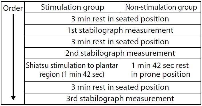
6.手順
事前に被験者には実験手順について十分な説明を行い、同意の上で行った。
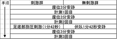
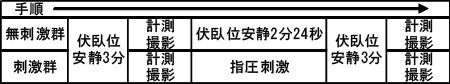
被験者に心拍計測用ディスポーサブル電極を装着し、口腔を洗浄するため水でうがいをさせ、肘かけ背もたれ付きの椅子に着席ののち5分間安静を保った。5分間経過後に1回目の唾液の採取と計測を行った(図1)。
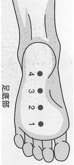
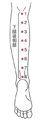
計測後、前頸部7)4点3回を左右ともに約1分間(1点約2秒×4点×3回×左右+移動時間≒ 60秒)快圧にて指圧した。指圧直後に2回目の唾液計測を行った。
2回目の計測後、更に5分間の安静後に3回目の唾液計測を行った。
7.記録
唾液アミラーゼの計測値はディスプレイに表示された数値をノートに記録するとともに、写真撮影による記録も並行して行った。
心拍数は、被検者に装着した電極からの信号をワイヤレスで受信した測定器と連動した PCに自動的に記録され、それぞれの唾液摂取時点の値を全測定終了後に抜き出してノートに記録した。
8.解析
唾液アミラーゼ、心拍数ともにうがい後5分間安静を保った時点の値を開始時点のデータとし、指圧直後および指圧5分後のデータ各間で対応あるt検定(Bonferroni補正)を行った。 有意水準は危険率5%未満(p<0.05)とした。
唾液アミラーゼと心拍数の相関についてはピアソンの相関係数を用いて判定した。
9.唾液アミラーゼ測定器について6)
NIPRO社製唾液アミラーゼ測定器(商品名:唾液アミラーゼモニター)はテストストリップ(商品名:チップ)を用いて採取した唾液を、機器本体に挿入し一定の操作をすることでテストストリップに内蔵された2種類の媒体、即ち唾液採取紙とアミラーゼ試験紙によって反応させたのち、アミラーゼを数値化して表示する仕組みである。交感神経の活動が活発になると分泌が促されるとされ、近年は比較的短時間のストレス指標としても用いられている。
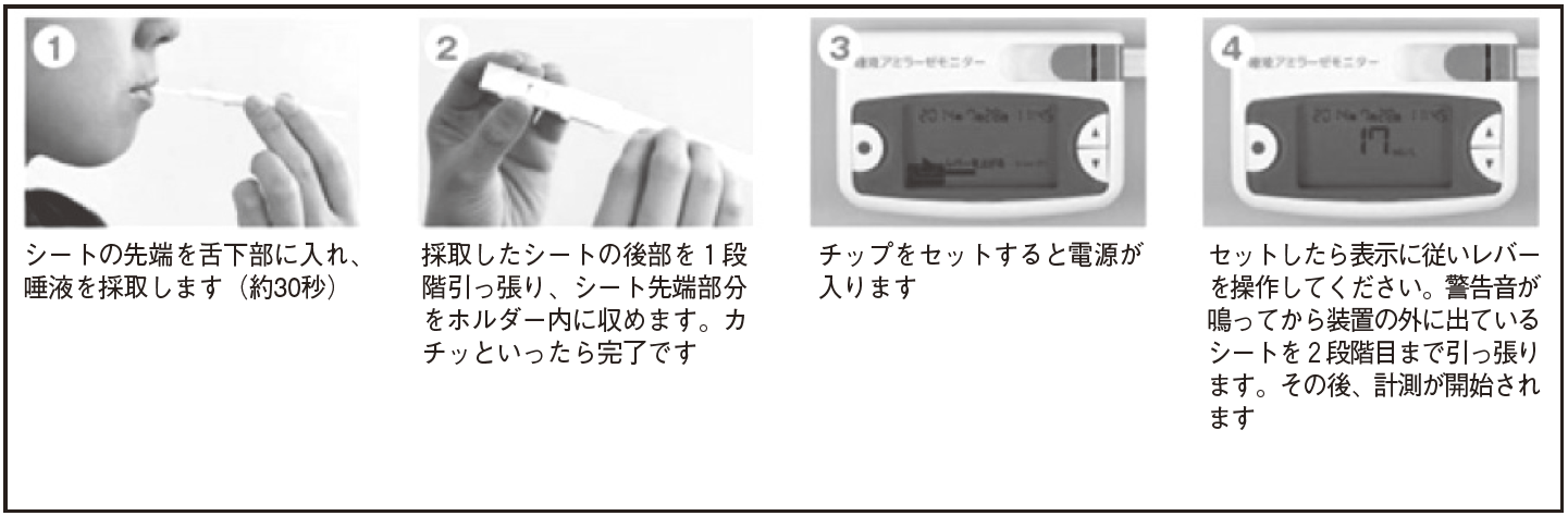
図1 唾液アミラーゼモニター使用方法8)
Ⅲ 結果
唾液アミラーゼ
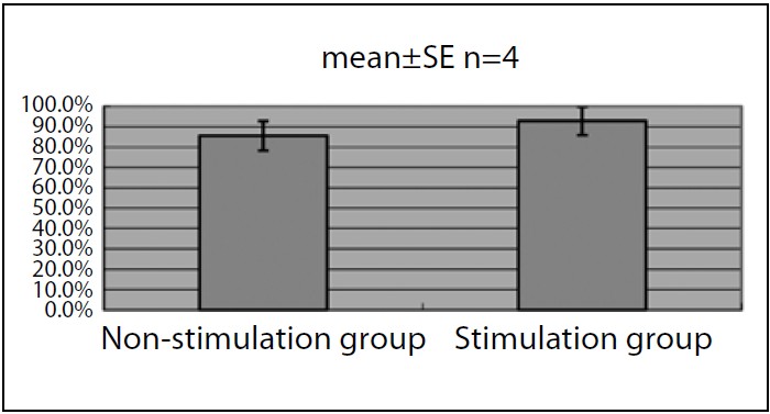
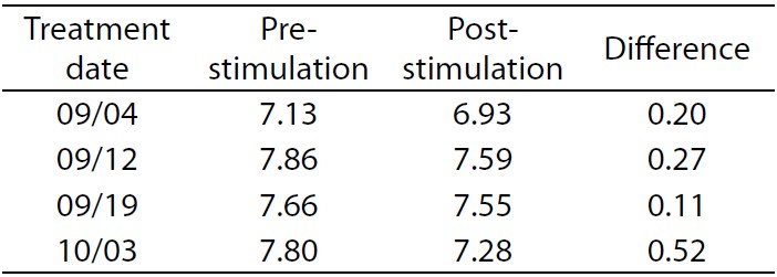
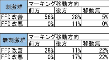
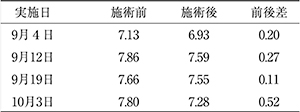
安静後と指圧直後では12名中7名が低下し、4名が上昇し、1名が変化なしであった。低下した7名中指圧5分後も引き続き低下したのは3名で、3名が上昇し、1名は変化がなかった。安静後と指圧直後で上昇した4名は、指圧5分後には全員が低下し、変化なしの 1名は上昇した(表1)。
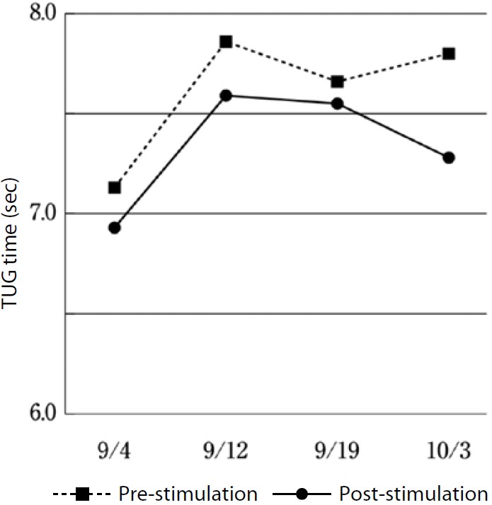
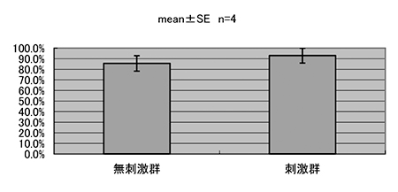
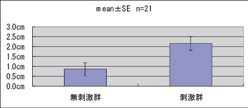
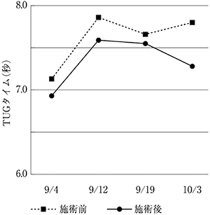
安静後と指圧直後では有意差は認められなかった(p=0.067)が、安静後と指圧5分後では有意差が認められた(p=0.016)(図2)。
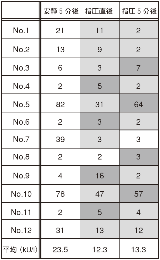
表1 唾液アミラーゼの変化
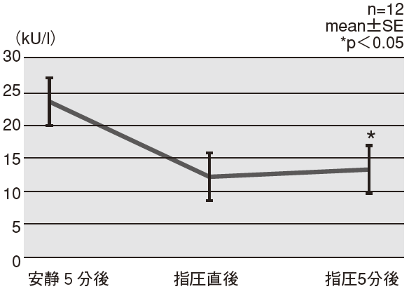
図2 唾液アミラーゼの変化
心拍数
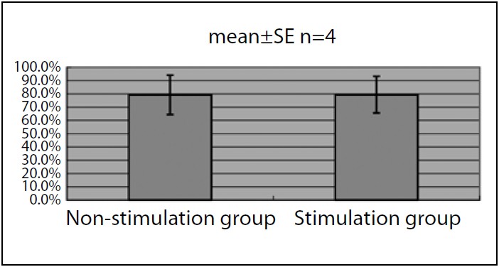
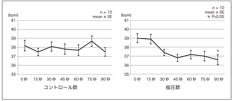
安静後と指圧直後では 12名中 11名が低下し、1名が上昇した。低下 11名中引き続き5分後も減少したのは 4名で、7名は上昇した。最初に上昇した 1名は、指圧 5分後では変化が無かった(表2)。
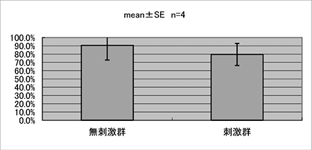
安静後と指圧直後で有意差が認められ(p=0.0001)、安静後と指圧5分後でも有意差が認められた(p=0.0008)(図3)。唾液アミラーゼと心拍数の間に相関はみられなかった。

表2 心拍数の変化
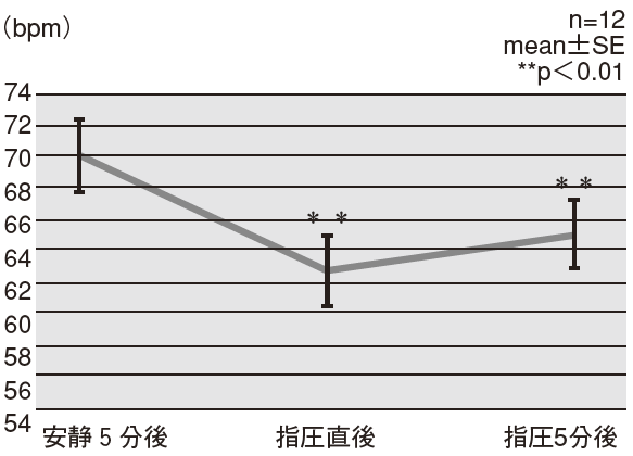
図3 心拍数の変化
Ⅳ 考察
前頸部指圧により心拍数が有意に低下する事象はこれまでにも報告され、今回も同様の成績を得られたことから再現性の高い現象であることが確認された。その機序については前報5)同様である。前報では心拍数低下に続いて HPA軸の亢進が起こる可能性を仮説として示したが、今回、唾液アミラーゼの値は全例が一様には上昇しなかった。安静5分後の初期値が比較的高い者は低下し、比較的低い者は上昇する傾向が観察された。機序として、指圧により初期値の高い者は延髄の上・下唾液核9)が刺激され副交感神経経由の唾液分泌量が上昇し、初期値の低い者は頸部交感神経9)が刺激されて唾液アミラーゼが上昇したと推察されるが、詳細は不明である。結果的には前頸部指圧により全体として指圧5分後には安静5分後より有意に低下し、ストレスが軽減した状態となった。
機序は不明であるが、指圧が HPA軸や他の内分泌のバランサーのような機構を亢進させていると仮定する。今回のように対象が健康成人である場合、各個人の初期状態にはばらつきがあると考えられるため、指圧による反応にもばらつきが生じたものと推察する。しかしながら、心拍数に関しては全例ほぼ一様に低下の反応を示し、加えて、前頸部指圧により呼吸商が低下する現象4)も今回とほぼ同率且つ同程度の時間経過で反応していることから、幾つかのシンプルな一次的反射を起点として二次的な反応が体内で引き起こされている可能性が考えられる。これは前頸部指圧により血圧が一過性に上昇したのちに血圧と心拍数が低下する現象2)とも相似すると考えるが、時間差を含めた詳しい機序の解明は今後の課題としたい。
加えて、唾液アミラーゼ分泌が有意に低下する時点は心拍数が有意に低下する時点よりも遅かったことは、浪越式基本指圧に於いて前頸部への施術が他の部位に先んじて行われなければならない一因であると推察する。
対象の初期状態に関しては、同一の病名を診断された複数の患者は初期状態が同程度の対象群であるとも換言でき、指圧治療による症状改善の報告がより一層多くなされれば、基礎研究レベルでは観察し得ない現象の考察が可能になるはずである。よって更に多くの症例報告を求め、機序解明の糧としたい。
Ⅴ 結論
前頸部指圧によって心拍数は指圧直後・指圧5分後ともに有意に減少した。唾液アミラーゼの分泌は指圧直後には有意差が無いが、指圧5分後には有意に減少した。
引用・参考文献
1)小谷田作夫 他:指圧刺激による心循環系に及ぼす効果について,東洋療法学校協会学会誌(22);p.40-45,1998
2)井出ゆかり 他:血圧に及ぼす指圧刺激の効果,東洋療法学校協会学会誌(23);p.51-56,1999
3)加藤良 他:前頚部指圧が自律神経機能に及ぼす効果 ,東洋療法学校協会学会誌(32);p.75-79, 2008
4)衞藤友親 他:前頚部指圧による呼吸商の変化 ,日本指圧学会誌(1);p.11-13,2012
5)衞藤友親 :ウマを対象とした前頚部指圧による心拍数の変化 ,日本指圧学会誌(3);p.16-18,2014
6)山口昌樹 他 :唾液アミラーゼ式交感神経モニタの基礎的性能 ,生体医工学 Vol.45 No.2;p.161-168, 2007
7)石塚寛 :指圧療法学 改訂第 1版,国際医学出版 , p.42,p70,2010
8)NIPRO:医療機器情報 http://med.nipro.co.jp/ index
9)佐藤優子,佐藤昭夫 ,山口雄三 :生理学 第 1版 15刷,医歯薬出版,p.58-59,2001
【要旨】
前頸部指圧による抗ストレス作用―唾液アミラーゼの変化より考察―
衞藤 友親
先行研究にて前頸部指圧が抗ストレスホルモンの分泌を促している可能性を示したのを受け、健康成人 12名を対象に前頸部指圧による唾液アミラーゼと心拍数を計測した。仮説に反し唾液アミラーゼ分泌量は上昇例も低下例も観察され、心拍数の低下傾向との相関はみられなかったが、両指標とも指圧5分後には有意に低下した。前頸部指圧によりストレスが低下する可能性が示唆される成績が得られたと考える。
キーワード:指圧、前頸部、唾液アミラーゼ、心拍数、ストレス